
Neuronavigation-Guided Keyhole Frontal Craniotomy for Tumor Decompression in a Patient with a Ring-Enhancing Frontal Lesion: A Case Report
Keyhole craniotomy has evolved as a minimally invasive neurosurgical technique that aims to provide adequate surgical access through a small opening while reducing surrounding tissue trauma, bony exposure, and morbidity related to the surgical approach. In appropriately selected cases, such as frontal tumors, this approach may achieve effective resection or decompression with favorable neurological and recovery outcomes.
In suspected high-grade glioma cases, the principle of surgical management is maximal safe resection, as an increased extent of resection has been associated with improved outcomes in appropriately selected cases. At the same time, contemporary perioperative pathways, such as enhanced recovery after surgery, have supported earlier mobilization and shorter hospital stays following surgery for brain tumors.
Case Presentation
A 63-year-old male presented with headache and involuntary movements in the form of generalized tonic-clonic seizures, followed by an episode of loss of consciousness. His Glasgow Coma Scale (GCS) score was E3V4M6 on repeated verbal stimulation.
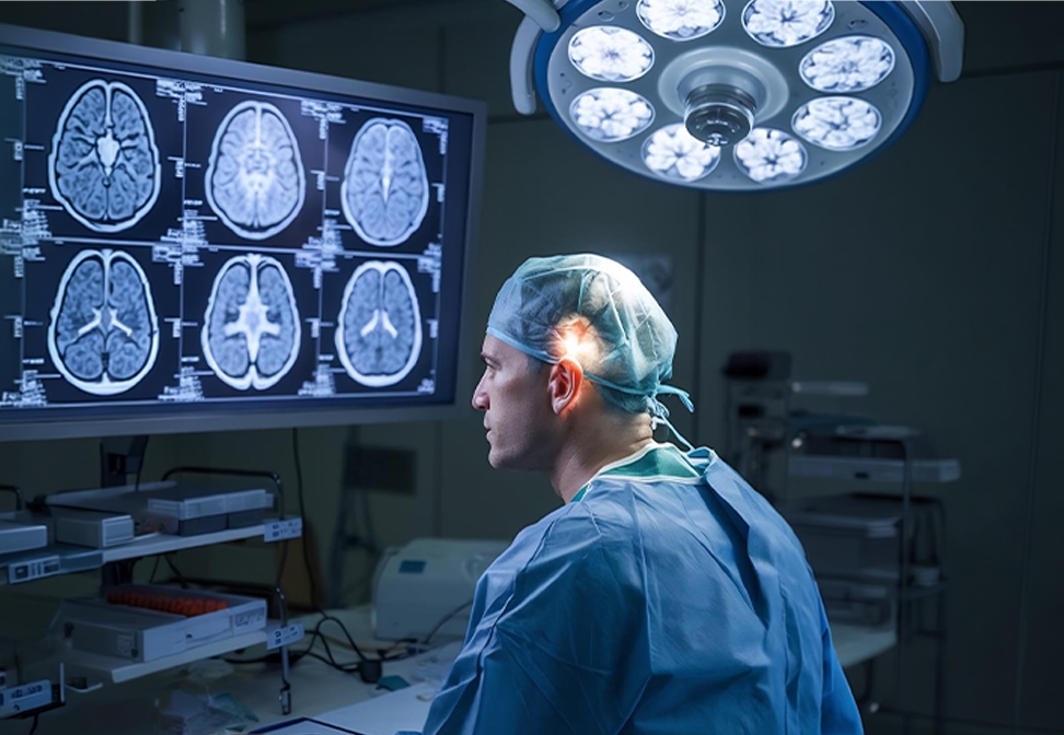
CT and contrast-enhanced MRI of the brain demonstrated a ring-enhancing frontal lesion with significant mass effect, raising strong radiological suspicion of a high-grade glioma. In view of the imaging characteristics, clinical deterioration, and compressive effect of the lesion, surgical decompression was planned.
Surgical Management
The patient underwent a neuronavigation-guided keyhole frontal craniotomy with tumor decompression. The scalp incision measured approximately 5 cm, and a 2.5-cm craniotomy was performed to provide a small, focused operative corridor to the lesion. Intraoperative neuronavigation was used to accurately localize the lesion and optimize the surgical trajectory, thereby minimizing cortical exposure and limiting the craniotomy size.
The postoperative course was smooth and uneventful. The patient was extubated in the immediate postoperative period, shifted to the room on the following day, and discharged on the second postoperative day. His neurological status improved after surgery, with a postoperative GCS of E4V5M6. This early postoperative recovery suggests that minimally invasive cranial approaches and structured perioperative recovery pathways may facilitate a reduced length of hospital stay in selected brain tumor cases.
Discussion
This case highlights the usefulness of a keyhole approach in combination with neuronavigation in the management of an aggressive frontal lesion producing mass effect and impaired consciousness. The keyhole concept is based on carefully planned, target-oriented exposure that provides sufficient working space while reducing collateral tissue damage. Reports on frontal glioma surgery have shown that keyhole approaches assisted by neuronavigation can be safe and technically feasible when patient selection, anatomical familiarity, and trajectory planning are meticulous.
The role of neuronavigation is particularly important in this setting because small craniotomies require accurate localization and precise surgical planning. For intrinsic frontal tumors, neuronavigation permits the surgeon to align the craniotomy and cortical access with the long axis of the tumor, thereby maximizing decompression through a small corridor.
Another notable feature of this case is the rapid postoperative recovery and early discharge. Recent literature on enhanced recovery following brain tumor craniotomy suggests that, with appropriate perioperative patient selection and workflow optimization, shorter hospital stays can be achieved without compromising safety. This case supports the view that minimally invasive cranial tumor surgery may contribute to improved postoperative recovery when applied judiciously.
A limitation of this report is that it describes a single case, with histopathological details and molecular markers pending. Nevertheless, it demonstrates the practical usefulness of a neuronavigation-guided keyhole approach in achieving effective decompression with satisfactory short-term neurological recovery in a patient with a lesion radiologically suggestive of a high-grade glioma.
Conclusion
Neuronavigation-guided keyhole frontal craniotomy is a useful minimally invasive option for selected cerebral intra-axial lesions with mass effect, including those radiologically suggestive of high-grade glioma. In this case, the approach allowed effective tumor decompression through a small craniotomy, with postoperative neurological improvement and discharge on the second postoperative day.