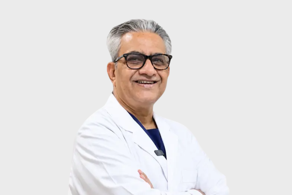
A rare and complex cardiac condition involving active infective endocarditis with intracardiac destruction was successfully managed under the leadership of Dr. Raju Vyas and the surgical and critical care team at Ujala Cygnus Hospitals.
The case involved the development of a Left Ventricular Outflow Tract (LVOT) to Left Atrium (LA) fistula, a challenging complication associated with significant structural damage to the heart. Such cases require meticulous surgical planning and precise reconstruction because of the involvement of critical cardiac structures.
A Complex Cardiac Condition
Infective endocarditis is an infection affecting the inner lining of the heart and, in severe cases, can cause extensive destruction of cardiac tissues.
In this case, the infection resulted in intracardiac tissue destruction and the formation of an abnormal communication between the LVOT and the left atrium.
The LVOT is the pathway through which blood leaves the left ventricle, while the left atrium receives oxygen-rich blood from the lungs. The presence of a fistulous connection between these chambers represents a complex structural abnormality requiring specialised surgical intervention.
The case was particularly challenging because of the extensive tissue involvement and the need to remove infected tissue while simultaneously restoring the affected cardiac anatomy.
Meticulous Surgical Reconstruction
The surgical team performed radical debridement, removing infected and necrotic tissue and preparing the affected area for reconstruction.
Following debridement, bovine pericardium was used for reconstruction of the cardiac defect.
The patch provided a flexible barrier to bridge the defect between the LVOT and left atrium and facilitate restoration of the affected anatomy.
The procedure also involved implantation of a tissue prosthetic valve.The combination of extensive debridement, anatomical reconstruction and valve replacement required careful surgical execution because of the complexity of the affected cardiac structures.
Encouraging 1-Month Outcome
The patient’s 1-month post-operative assessment demonstrated a stable clinical status, with echocardiographic evaluation showing complete resolution of the intracardiac shunt.
The tissue prosthetic valve also demonstrated a single digit mean transvalvular gradient, indicating satisfactory early prosthetic valve haemodynamics.
This early outcome reflects the successful restoration of the affected cardiac anatomy following a highly complex surgical reconstruction.
Key Surgical Considerations
Radical Debridement In destructive infective endocarditis, complete removal of infected and necrotic tissue is an important component of surgical management. The surgical objective is to establish healthy tissue margins before proceeding with reconstruction.
Bovine Pericardial Reconstruction
A bovine pericardial patch can be used to bridge structural defects and reconstruct affected cardiac tissue. In this case, it was used to address the defect involving the LVOT and left atrium.
Tissue Prosthetic Valve
A tissue prosthetic valve was implanted as part of the reconstruction. At the 1-month follow-up, the valve demonstrated a single-digit mean transvalvular gradient, indicating satisfactory early haemodynamic performance.
Follow-Up After Complex Cardiac Reconstruction Long-term follow-up is important following complex infective endocarditis surgery and intracardiac reconstruction.
The clinical document recommends ongoing echocardiographic surveillance to monitor the stability of the reconstructed area, assess prosthetic valve performance and identify potential structural complications such as pseudoaneurysm formation.
Monitoring for recurrence of infection and appropriate infective endocarditis prevention measures are also important components of post-operative care. The source recommends monitoring inflammatory markers such as ESR and CRP until baseline normalisation.
A Successful Outcome in a Rare and Challenging Case
The successful management of this complex case highlights the importance of specialised cardiac surgical expertise, meticulous removal of infected tissue and precise anatomical reconstruction.
At the 1-month post-operative assessment, the patient remained stable, with complete resolution of the LVOT-to-LA intracardiac shunt and satisfactory early tissue prosthetic valve function.
The successful outcome represents an encouraging result in the management of a rare and technically demanding cardiac condition.